How is 3D mammography different from 2D mammography?
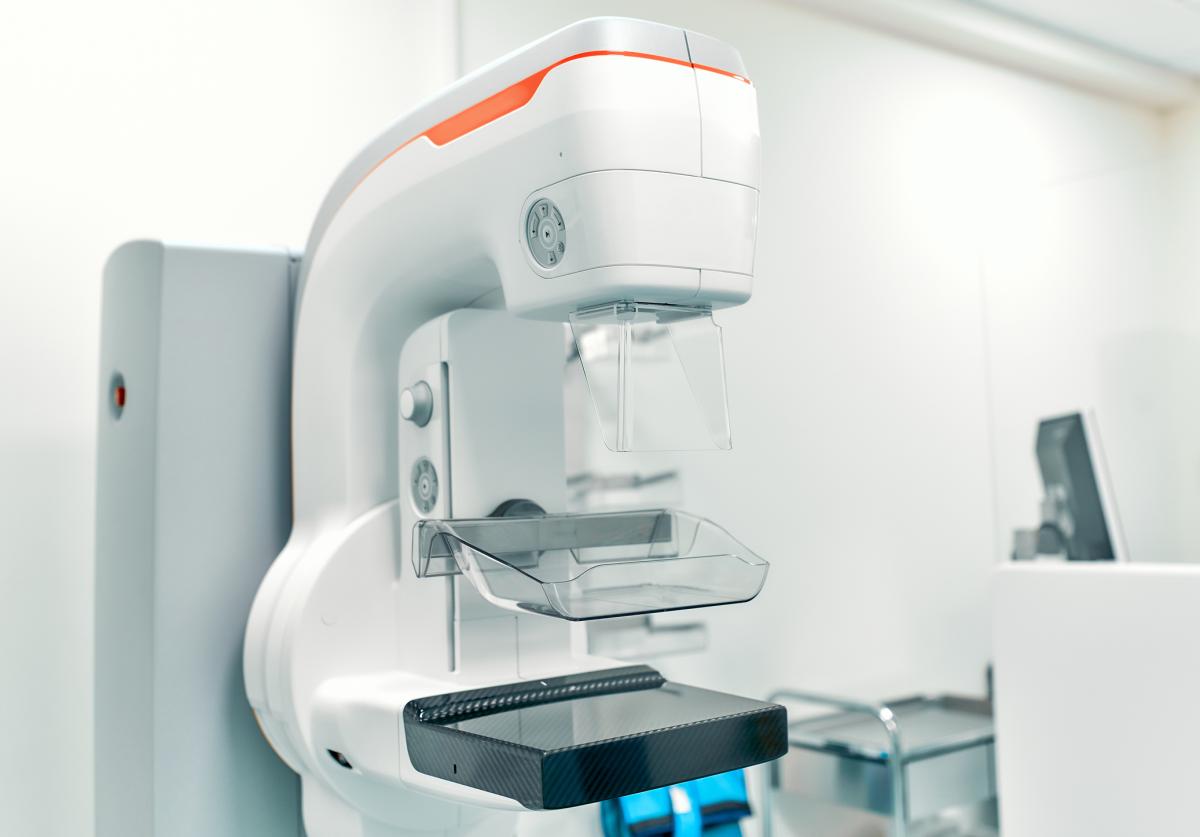
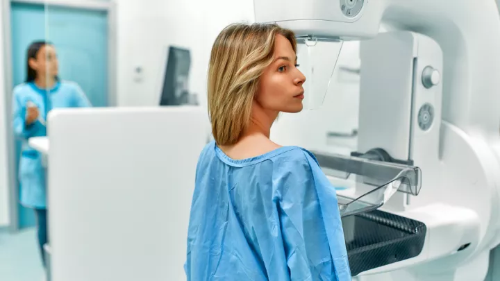
The 3D mammography screening experience is similar to a traditional mammogram. During a 3D mammography exam, multiple, low-dose images of the breast are acquired at different angles. These images are then used to produce a series of one-millimeter thick slices that can be viewed as a 3D reconstruction of the breast.
Research shows breast cancer screening with 3D mammography, when combined with a conventional 2D mammography, has a 40% higher invasive cancer detection rate than conventional 2D mammography alone. It also provides a significant reduction in “call back” rates of 20% to 40%.

“In large studies, it’s been proven that more cancers were found and more cancers on a smaller stage were found — in all levels of density of the breast,” says Cheryl Williams, MD, radiologist. “This is very important. The smaller a tumor is when we find it, the more likely it is that we’ll be able to cure it.”
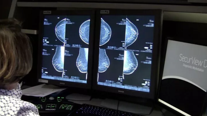
How does it actually work? The system uses high-powered computing to convert digital breast images into a stack of very thin layers, or “slices” – building what is essentially a 3D mammogram. This allows radiologists to examine the breast tissue one layer at a time, without the confusion of overlapping tissue.
When the overlapping tissues within the breast are viewed as a 2D, flat image, it can become confusing to radiologists reading the image. This is the leading reason why small breast cancers may be missed and normal tissue may appear abnormal, leading to unnecessary call backs.
Breast cancer is the second leading cause of cancer death among women, exceeded only by lung cancer. Statistics indicate one in eight women will develop breast cancer sometime in her lifetime. The stage at which breast cancer is detected influences a woman’s chance of survival. If detected early, the five-year survival rate is 91%.
“Bottom line, get a mammogram.” Dr. Williams says. “Get it every year after the age of 40. Be consistent. It truly is a life saver.”


