New thoracic branch graft procedure improves outcomes for patients with aortic tears
Michael Stephens retired several years ago, but you’d never know it. The 73-year-old from Loup City, Nebraska, likes to keep busy. If he’s not helping friends with farm work, he’s out clearing land, trimming trees or operating other large equipment.
Several years ago, however, chest pain landed him at the hospital, where Stephens was diagnosed with an aortic dissection, a tear in the largest blood vessel of the heart. The tear was not serious enough for surgery, so following protocol, Stephens was put on a blood pressure- lowering medication and monitored with frequent CT scans.
He was advised to slow down and avoid lifting items more than 5 to 10 pounds or risk increasing the tear. Slowing down was not what Stephens wanted to hear, but he reluctantly abided as well as he could.
Earlier this year, Stephens got the news that the tear had increased and the aneurysm (thinning and enlarging of the aorta) had grown.
“Michael had developed a third lumen or tube in the artery, the most dangerous situation,” says Jason Cook, MD, Nebraska Medicine vascular surgeon. “We recommended surgery to repair the aorta and reduce his risk of rupture and dying.”
Today, Stephens is back to doing the things he loves with no restrictions, thanks to a new procedure now performed at Nebraska Medical Center called a thoracic branch graft.
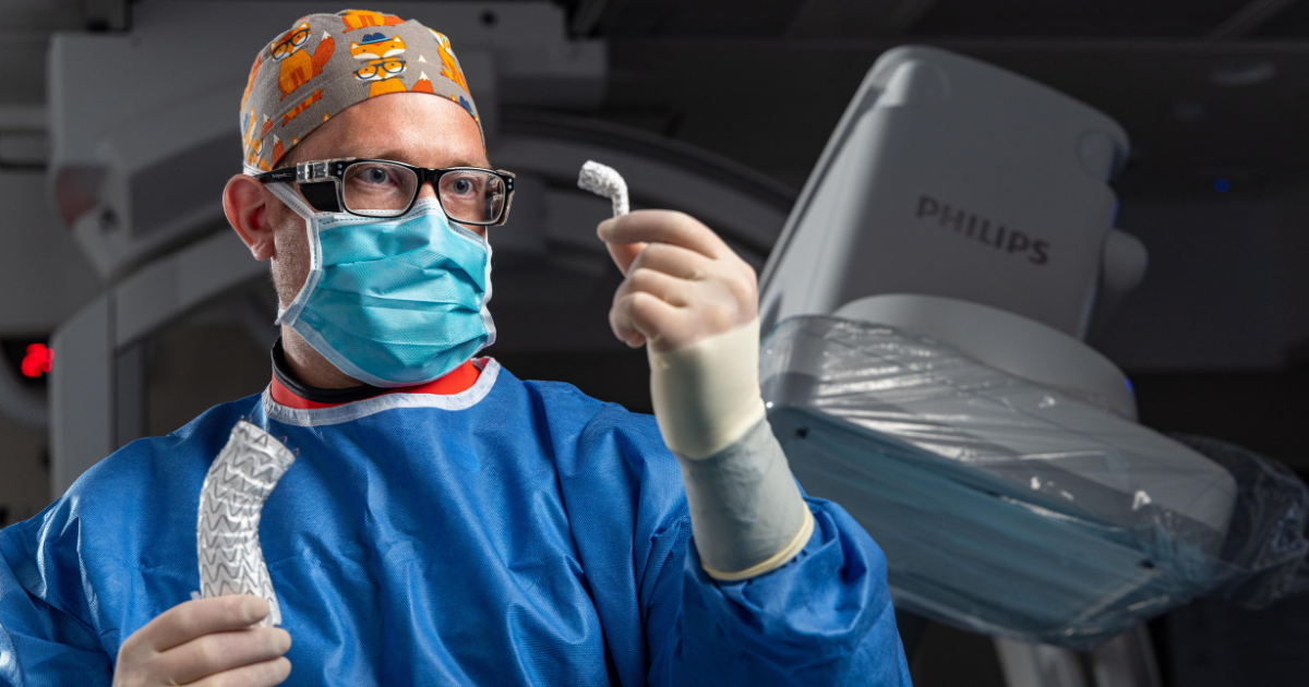
The minimally invasive procedure requires only two small needle sticks in the groin and left arm. A catheter containing the stent graft is inserted and guided through the artery to the diseased aorta and carefully positioned to seal off the damaged area.
The aorta is the largest blood vessel in the body. It carries blood from the heart to the rest of the body through smaller branched arteries. This procedure uses a new stent graft that includes a portal that lines up with the left arm artery to prevent issues with blood flow to the left arm. When properly aligned, it allows for continued blood flow to the smaller branched arteries.
Before this new device became available, doctors had two options. They could perform open chest surgery to repair the damaged aorta, or they could perform two surgeries. The first open surgery required a bypass from the left neck to the left arm, followed by a second surgery to seal off the damaged segment of the aorta with a stent graft delivered through the groin.
“This is a significant improvement in the evolution of patient care with aortic disease, including aneurysms and dissections,” says Jason Cook, MD, Nebraska Medicine vascular surgeon. “When positioned precisely, it is extremely effective with over a 95% success rate in clinical trials.”
The procedure requires no incisions and eliminates the risks associated with open chest and bypass surgeries. In addition, Dr. Cook notes that the patient can typically go home within 48 to 72 hours and resume normal activity within a week.
“I’m really glad I had the procedure done,” says Stephens, as he prepared to start a day of work at a friend’s farm. “I like to be doing something. I don’t like being a couch potato.”


